Health>Mind+Body: Herbs we sell: Listed below are Herbs i sell and endorse from there affiliate program The King of Medicinal Mushrooms, according to Sib...
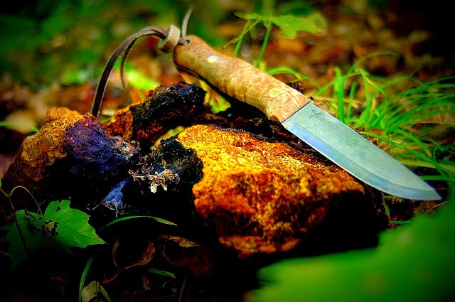
CHAGA: King of the Medicinal Mushrooms
Saturday, January 3, 2015
Friday, July 25, 2014
sunscreen
Wear sunscreen That may not be the best advice for everyone all the time

How is that a problem? It turns out that a lack of sun exposure can cause serious health problems for some people, and my doctor says I am one of them.
Studies show that people with dark skin, such as myself, rarely get skin cancer no matter how much exposure to the sun we get. When we are afflicted, it’s usually in spots on the body that rarely see daylight, such as the bottoms of feet or between fingers, also places one rarely slathers the SPF50.
Our bodies manufacture a chemical called melanin. It’s responsible for pigment, and there are two varieties that determine how brown we are. Owners of the first kind have dark skin, tan in the sun and rarely burn. Let’s call this one Lisa melanin. The other type is found in people who are pale, rarely tan and burn in the sun. We’ll call this one Copier Paper. Just kidding. We’ll call this one Hubby melanin, after my color-free beloved.
Melanin has another very important role besides pigment. It helps our bodies make Vitamin D. The only way humans can manufacture Vitamin D, necessary for everything from strong bones to immune health, is by exposing our skin to the sun. And humans bathing in sunscreen or lost in the cubicle labyrinth aren’t subjected to sunlight. No sun, no home-brewed Vitamin D.
For people with Lisa melanin, not having sun exposure is worse than for people with the other version. The Hubby kind, which evolved in Northern Europeans and the people from the British Isles, helps make Vitamin D six to 10 times faster than the original, found in people whose origins are in sunny climes such as Africa and India.
So people like my husband need about five to 15 minutes of sunlight a few times a week. And people like me need between 30 and 150 minutes — Coppertone-free.
The CDC makes no distinction. It advises Lisas to use sunscreen, period. Then they show up in their doctors’ offices severely Vitamin D-deficient. Next, they find themselves popping Vitamin D megadoses to ward off bone damage and immune problems. (This Lisa’s prescription is from her rheumatologist.)
Not to worry, says the CDC, the American Academy of Dermatology, and every American federal bureau and organization of muckety-mucks. Slam down those supplements or snack on cod liver oil capsules and you’ll be fine. Just don’t soak up any rays.
Melanin science is complex and controversial. As investigative journalist Jessica Seigel documents in June’s edition of Nautilus, an eminent professor and foremost expert on Vitamin D was fired for suggesting that Americans need some sun. He had proposed that humans are perfectly fine spending about five minutes sunning themselves before basting with sunscreen.
While Americans are scandalized by that radical idea, the Australian government and cancer councils have been making the same recommendations for the last decade. Keep in mind that Australia is the skin cancer capital of the world. Aussies are expected to check the daily UV index. If the sun’s ultraviolet rays are low enough, a 3 or less on the scale, then no one — not even Hubby needs sunscreen. The SunSmart campaign even has an app for that.
Official U.S. policy discourages Americans from creating their own Vitamin D and encourages us to take supplements at doses that are much lower than many experts recommend. By saving us from skin cancer, our government means well. By downplaying our Vitamin D needs, it could be killing some of us with kindness.
source
http://www.dallasnews.com/opinion/latest-columns/20140725-wear-sunscreen.-that-may-not-be-the-best-advice-for-everyone-at-all-times..ece
Tuesday, July 22, 2014
sunburn+tanning activators
Tanning activators are chemicals that increase the effect of UV-radiation on the human skin.
Since sunburn and suntan are induced by the same mechanism direct DNA damage, these substances increase the likelihood for sunburn as well. The best known tanning activator is psoralen which is an ingredient of bergamot oil. Psoralen has been present in sunscreens in order to allow suntanning despite the reduced UV-intensity that acts on the deeper layers of the skin. In Switzerland, a ban was imposed on psoralen containing sunscreens in 1987 but it was loosely enforced for several years. In other countries these substances have been present in sunscreens until the first epidemiological results have shown that users have a fourfold risk of developing melanoma. They were finally banned in 1996. This happened more than 15 years after the photocarcinogenic potential of psoralen had been demonstratedAfter the evidence for the photocarcinogenic potential of psoralen emerged, sunscreens which combined UVB filters and psoralen were introduced onto the market. These products were accompanied by campaigns to convince the public and the regulatory authorities that these products were safe or even better than usual sunscreens. These sunscreens were especially recommended to poor tanners. Psoralen tanning lotions were available in France, Belgium and Greece.
The increased production of melanin is the reaction of the skin to UVB-induced direct DNA damage. Several substances are known to increase the amount of direct DNA damage thymine dimers. In order to produce this action they have to penetrate into the skin, and this is in contrast to the assumptions which are made by those who endorse sunscreen use .
The tanning activator coumarin is known to induce thymine dimers (cyclobutane pyrimidine dimers).
Other Web sites state correctly that: "Coumarins produce photosensitivity therefore advise the patient to avoid direct sunlight after treatment.
Melanocytes melanin
Melanocytes are melanin-producing cells located in the bottom layer the stratum basale of the skin's epidermis, the middle layer of the eye the uvea, the inner ear, meninges, bones, and heart.Melanin is the pigment primarily responsible for skin color.

Through a process called melanogenesis, these cells produce melanin, which is a pigment found in the skin, eyes, and hair. This melanogenesis leads to a long-lasting pigmentation, which is in contrast to the pigmentation that originates from oxidation of already-existing melanin.
There are both basal and activated levels of melanogenesis; in general, lighter-skinned people have low basal levels of melanogenesis. Exposure to UV-B radiation causes an increased melanogenesis. The purpose of the melanogenesis is to protect the hypodermis, the layer under the skin, from the UV-B light that can damage it (DNA photodamage). The color of the melanin is dark, allowing it to absorb a majority of the UV-B light and block it from passing through this skin layer.
Since the action spectrum of sunburn and melanogenesis are virtually identical, they are assumed to be induced by the same mechanism. The agreement of the action spectrum with the absorption spectrum of DNA points towards the formation of cyclobutane pyrimidine dimers (CPDs) - direct DNA damage.
Typically, between 1000 and 2000 melanocytes per square millimeter of skin are found. Melanocytes comprise from 5% to 10% of the cells in the basal layer of epidermis. Although their size can vary, melanocytes are typically 7 μm in length.
The difference in skin color between lightly and darkly pigmented individuals is due not to the number (quantity) of melanocytes in their skin, but to the melanocytes' level of activity (quantity and relative amounts of eumelanin andpheomelanin). This process is under hormonal control, including the MSH and ACTH peptides that are produced from the precursor proopiomelanocortin.
Albinos lack an enzyme called tyrosinase. Tyrosinase is required for melanocytes to produce melanin from the amino acidtyrosine.
Numerous stimuli are able to alter melanogenesis, or the production of melanin by cultured melanocytes, although the method by which it works is not fully understood. Melanocortins have been discussed to have effect on appetite and sexual activity in mice. Vitamin D metabolites, retinoids, melanocyte-stimulating hormone, forskolin, cholera toxin,isobutylmethylxanthine, diacylglycerol analogues, and UV irradiation all trigger melanogenesis and, in turn, pigmentation.Increased melanin production is seen in conditions where ACTH is elevated, such as Cushing's disease. This is mainly a consequence of alpha-MSH being secreted along with ACTH. Alpha-MSH is a cleavage product of ACTH that has a stronger affinity for the MC1 receptor on melanocytes.
Once synthesised, melanin is contained in a special organelle called a melanosome and moved along arm-like structures called dendrites, so as to reach the keratinocytes. Melanosomes are vesicles which package the chemical inside a plasma membrane. The melanosomes are organized as a cap protecting the nucleus of the keratinocyte.
When ultraviolet rays penetrate the skin and damage DNA, thymidine dinucleotide (pTpT) fragments from damaged DNAwill trigger melanogenesis and cause the melanocyte to produce melanosomes, which are then transferred by dendrite to the top layer of keratinocytes.

Tuesday, July 15, 2014
Melanin
Melanin also forms stable free radicals, quenches excited states, and binds radical-forming agents such as transition-series metals. All likely contribute to its putative role in antioxidant defense. On the other hand, the ability of melanin to bind toxic radical-generating agents may sometimes be detrimental, as in chloroquine retinopathy and aminoglycoside ototoxicity (41). Finally, melanin can function as an efficient S0Dase and may retain this function in pigmented organs. Thus, the melanins ( which can form abiologically ) may be the oldest evolved system for defense against oxygen radicals, rather than SOD/catalase.
Free radicals are produced by environmental causes such as light or ionizing radiation. However, three physiological processes can result in extraordinarily high levels of radical species. These include the mixed-function oxidase system of endoplasmic reticulum, the NADPH oxidase system of inflammatory cells, and the presence of high levels of autoxidation-mediating charge-transfer agents. Production of activated species by such mechanisms can exceed the capacity of local protective mechanisms and produce tissue injury.
Inflammatory cells produce active species of oxygen in antimicrobial defense (1,2). While such species may directly damage surrounding tissues, their major secondary role may be to mediate important components of the inflammatory response. For example, Figure 3 lists some of the inflammatory immunomodulators reported to be affected in vitro by one or more components of the active oxygen system. Inflammation in the general sense comprises the whole of the systemic response to injury, so many of these same processes may also occur in ischemic injury, for example. While circumstantial, the list includes most of the major components of the inflammatory response and grows daily.
Similarly, antioxidants, SOD, and catalase have significant anti-inflammatory properties (3-5). For example, Orgotein, the pharmaceutical preparation of SOD, is used in veterinary medicine. It is reported to be both safe and effective in the treatment of various inflammatory and degenerative lesions in man ( 3-4 ). The action of many other antiinflamatory drugs may also involve interactions with the active oxygen system.4 Such agents may act by interfering with the action of phagocyte-produced active oxygen species on one or more of the systems outlined in Figure 3. The role of active species in the inflammatory response may also explain the dermal pigmentary response in inflammation (4). Active oxygen species may also have a role in endotoxin shock, burn-induced plasma volume loss, and even in atherosclerosis - e.g., the atherosclerotic lesions in homocystinuria. Likewise, radical mechanisms may play a role in stroke, cerebral edema, and spinal cord injury, as well as ischemic injury ( 42 ).
Physics and Chemistry of Melanins.
Physics and Chemistry of Melanins.
The peculiar characteristics of the melanins are always the colour and their free radical nature (6). Scarse results have also been found with the most sophisticated apparatus (X-rays, MS, NMR, Laser) (Link 22).
There are three fundamental theories on the chromophores of melanins. The first is that which proposes that melanin is a mixture of chromophores which resonate in different parts of the spectrum. A mixture of red, blue, green etc. chromophores absorb the radiation of all the wave lengths of visible light and appear black. Another theory predicts that a highly conjugated system produces black, brown and red-brown depending to the gap value of the semiconductors. What is the extension of the conjugation that allows a body to appear black? Many natural substances possess extended systems of conjugated orbitals (for example the carotinoids) but none of these compounds appears black. The study of the electronic structure of an ordered and ideal polymer of 5,6-indolquinone which is a common component of the eumelanins (hair, eyes, skin, etc.), conducted using the theory of Hückel, predicts that the black colour appears in a polymeric unit with a number of monomers higher than 10 and up to a infinite polymer and that, besides, it is an amorphous semiconductor and a good conductor of electricity (3) . The theoretical model is not supported by few experimental data. The only experimental data which currently exist for polyarene and polyene structures are those relative to pyrrole- black and acetylene -black which, as has been said, are radical-polarone system. ( Link 9, 12, 21, 22 ) In the study of the melanins it has often been claimed that the different colours, yellow, orange, red, red-brown, black are due to the type and the size of the granules of the pigment and to the distribution of the granules in the tissue. A third theory proposes that the melanins are schemochromes, that is that the colour depends on particular particle structure. The melanins could be a sort of black body in which the light which penetrates is reflected and diffused until it is completely absorbed. The cause which produces the "black" must be studied using solid state theory and band model of semiconductors.
Melanin as stable free radical .
Despite the fact that every chemical and physical study of melanin must be interpreted with more care compared to what would usually be the case in the study of pure and crystaline substances, the black melanins (eumelanins), brown, red-brown, and yellow melanins (pheomelanin), the allomelanins and the humic acids present a characteristic EPR signal, sometimes with some hint of a hyperfine structure (Link 22). The origin of the paramagnetism is still a controversial problem. Several efforts, (nothing is ever clear and definitive in scientific research on the black substances), have been made to correlate the free radical nature of the melanin with certain biological functions, like the physiology of vision, photoprotection, threshold switching, the electret effect etc. Studies have been carried out on all the melanins in acqueous suspensions and almost always give an EPR signal at about 4-6 G. The spin concentration is around the value 4-10 x 1017 spin/g. In the "polymer" there would be one free radical every 200-1,000 "monomers". It would seem that there are two radicalic centres in the black products that originate from the o.phenols: one being essential (intrinsic), highly stable, generated in the course of melanogenesis and "trapped" in the product and the another being extrinsic, transient and reactive which can form in the melanin by the action of the different chemico-physical agents. Passing from black melanins to brown and red-brown products (pheomelanins) it is possible to observe radicals with better defined structures, at different pH, like those of semiquinonamine and semiquinone. EPR studies carried out on the hair and skin of several bovine races and on albinoes have mainly been used by geneticists and pathologists. Albino subjects, with the same phenotype character, have hair with differing electronic characteristics. In some albino subjects there is a weak EPR signal which is completely absent in others. There are, that is, true albinoes and false albinoes (6, l).
The chemistry of free radicals, which has almost always been associated to the processes of polymerisation and oxidation, has developed in isolation from the context of organic chemistry. The chemistry and biochemistry of natural substances have always considered the free radicals with diffidence because of their reactivity which makes them difficult to control. It is probable that different radicalic reactions intervene in melanogenesis. Melanogenesis has never been considered as an essentiallly radicalic process. The EPR signal, present in all the melanins, has been attributed to a system like the cyaninic colourants (merocyanines) which give, as is known, EPR signals similar to those of the melanins even though they do not have unpaired electrons. This explanation of the meaning of the EPR signal by the merocyanines seems strange? The hypothesis that the signal comes from an inert radical of the copolymerisation material of the melanin cannot be accepted. A somewhat surprising observation is that the radical is little present in the melaninin granule and therefore of little significance , while it is highly probable that the important properties of the melanin (solubility, colour, reactivity, conductivity) are linked to this electron which lives alone in a gap of the granule. When the melanin lightens from black it becomes brown, red-brown, yellow, that is one passes to pheomelaninic granules (hair, fur, feathers, eyes), not only is the EPR signal always present but it appears in a more and more structured form, to the point that it is possible to distinguish the black melanin from the red-brown (pheomelanin). The EPR signal is present both in melanins prepared in the laboratory under most varied conditions and in pheomelanins of very varied origins. For the humic acids of the hydrosphere and of the lithosphere, so important for life, one has a very similar general picture even if the heterogeneousness of the material makes the granules of the humic acid much more difficult to study. The results obtained with IR, NMR, X-rays and MS cannot be believed significant. The various spectra which often present absorption characteristics, may not be due to the humic acid but to incorporated substances (which can be used for the qualitative and quantitative analysis of terrains and of waters). The EPR signal is pure, clear and very similar to that of the melanins. In general one obtains the EPR signal of two types of stable radical which must be attributed either to the pyrocatechine-resorcine type or the quinhydrone type. In general one has signals of various intensity according to the origin of the humic acid (humic acids from micro-organisms, soluble humic acids, black carbon humic acids, grey humic acids, brown humic acids, marshland, manure, peat). Notable paramagnetism is present in the fungal humic acid (Cephalosporium gordoni) which is also darker: the blacker the composite the more intense the EPR signal. It is calculated that for a concentration of 1017 spin/g in a "composite" of molecular weight 10,000 there is 1 radical for every 600 monomers. It is plausible that, from a structuralis chemical point of view, the part of the "macromolecule", perhaps the chromatic part (acetylene-black), responsible for the paramagnetic phenomenon is assimilable to that of the more typical melanins present in the animals or produced by micro-organisms. Therefore the history of the EPR signal is identical for all the melanins regardless of their origins. As often happens in research the failure of an objective (identification, for example, of the structure responsible for paramagnetism in the melanin) produces new and useful knowledge. The discovery that the humic acids of various origins and natures are free radicals has given useful information on all the existing relationships between the EPR signal and the properties of these substances. In the soil there are all the conditions for the genesis of free radicals: water, light, heat, organic and inorganic catalysts (Fenton reactive types). The soil is all a melting pot of radicals; in the soil the primordeal matrix can operate with ease .
The hypothetical radical melanogenes of the soil can form through the action of oxygen and light on products deriving from micro-organisms, from the oxidative demolition of the lignin, and from other biological polymers. In the physiology of plants humic acids act both as growth factors and as activators of cellular respiration. The semiquinonic radicals of the humic acids influence germination and, linking to atmospheric oxygen, transport active oxygen in the soil. The humic acids as polycations fix and transfer precious counteranions to the life of the soil and the waters.They can be good conductors when opportunely prepared and doped. In conclusion, polymers like melanin from DOPA, melanin from sepia, humic acids, melanin from phenols, like pyrocatechine and pyrrole blacks, etc. , apparently differing among themselves, exhibit an identical signal at EPR which can be reinforced up to even 100 times if the measurement is made on sodium salts. The melanins of the soil (humic acids) form by a fluctuating and non-reproducible mechanism where time must be considered as an intrinsic parameter of the dynamic of the natural process and where the activation of water is a fundamental parameter (also cellular liquid). The concept of time is a problem of fundamental importance for the biologist of melanin, in that in its genesis this is not free from environmental and cosmic forces and influences or from the activation of the water under the action of electromagnetic fields or from solar phenomena in general. To show that some reactions cannot be located in the current logic of science, Piccardi stressed that accepting time as a coordinate negates the fundamental dogma according to which only reproducible experiments are valid. In the case of melanin, in fact, it is not possible to control the conditions in which an experiment is conducted without taking into account the time coordinate, because during its course the conditions in which an experiment, a chemical or biochemical reaction, is conducted change. In experiments on melanin the time may not be considered an isotrope in every direction in space, nor homogeneous for every successive instant. Melanin cannot be represented by a formula, with a complete melanogenetic scheme in that it is not representable in terms of linear equations because it is not possible to hypothesise a correspondence between cause and effect in the temporal succession of the relationships. A negligible reaction in a fixed instant can become a determining reaction in a succeeding instant.
In melanogenesis many classical concepts, definitions, chemical dogmas are obsolete, they are lost in the past. If the experimentor looks at the scientific explanation of the natural processes and is not content with the measurements of the immediately measurable quantities then he understands that he is working in an imperfect way. The analysis (combustion) of the melanins coming from the sack of the sepia are non-reproducible even with the same method of preparation of samples coming from the same source. To the chemist the synthesis of black content in the sack of the sepia must seem a non-reproducible process and is perhaps fluctuating because of the interference of magnetic activity and radiation of the sun on the sea, of the atmospheric electrical potential, of the variations in the Earth’s magnetic field.
The spirit of research in the field of the melanins must be renewed, accepting the hypotheses works some researchers, sometimes students, most of the time young emarginated researchers, would like to introduce into the world of research.
Melanin and skin cancer Again
Melanin and skin cancer Again
A new study by University of Kentucky researchers shows how a genetic defect in a specific hormonal pathway may make people more susceptible to developing melanoma, the deadliest type of skin cancer.

Fair-skinned people who tend to burn (rather than tan) from sun exposure have a much higher risk for melanoma than darker-skinned people. On the surface, it appears that the amount of melanin, the natural substance in the skin that determines pigment and acts as the skin's "natural sunscreen," would be the only determinant of melanoma risk. However, the truth is more complicated.
Published in Molecular Cell, the study looked at the role of the melanocortin1 receptor (MC1R), the receptor on melanocytes in the skin that gets called into action following ultraviolet exposure to help the skin lay down more UV-blocking melanin to protect itself. Fair-skinned people are more likely to inherit a defect in this receptor, and as a result, cannot make enough melanin to fully protect themselves from UV damage.
Since UV from sunlight or tanning beds is a major cause of melanoma, inherited problems in the MC1R means that the skin lacks natural protection by melanin, which acts as a biologic sunblock. This leads to more UV light chronically getting through to the sensitive layers of the epidermis, where it can contribute to cancer.
However, the UK study showed that MC1R defects contribute to melanoma development in ways other than melanin production. Besides regulating the amount of melanin that gets made in the skin, MC1R also controls how well melanocytes can repair their DNA from UV damage. Having defects in MC1R signaling delays the body's ability to clear out existing DNA damage in the skin – leading to an increased potential for cancerous mutations.
"Knowing whether people have a specific genetic predisposition for melanoma could potentially save many lives", says Dr. John D'Orazio, Associate Professor and the Drury Pediatric Research Endowed Chair at UK's Markey Cancer Center. "If you happen to be born with a problem in this MC1R hormonal pathway, then you need to be extra careful with respect to UV safety."
A good indication of a person's MC1R status is what happens to the skin after sun exposure.
"If you tan well, then your MC1R probably works well," D'Orazio said. "If you tend to burn, then you may have inherited a problem with your MC1R, and you probably should avoid purposeful UV exposure like tanning bed use or unprotected sun exposure."
D'Orazio and his research team found an important molecular link between MC1R signaling and DNA repair in their study. The team hopes to use this information to develop new melanoma-preventive treatments, like additives that can be included in sunblocks to ramp up the skin's ability to deal with UV damage.
Melanoma incidence has increased steadily over the past few decades – in the 1930s, an estimated one in every 1,500 Americans developed the diseases. Today, the odds are about one in every 60. Having a problem with the MC1R pathway raises a person's lifetime risk of melanoma about four-fold.(1)
Order Here .......>Chaga Great Video on Chaga
a great article on Chaga and article Quest for Chaga
Melanin and skin cancer Again#melanin + #sunburn
Subscribe to:
Posts (Atom)